Biochemistry of Medicinals I Phar 6151 CHAPTER TWO

Instructor: Dr. Natalia Tretyakova, Ph.D.
PDB reference correction and design Dr.chem., Ph.D. Aris Kaksis, Associate Professor

 
DNA Synthesis
A. DNA Polymerases
 
The synthesis of DNA is catalyzed by DNA polymerases.  They function by catalyzing the step-by-step addition of deoxy-ribo-nucleotides to the 3' end of a DNA polymer chain.  These enzymes are important for the error-free duplication of the DNA duplex, thus maintaining the fidelity of genetic information.
 
1. Polymerization Reaction
 
DNA polymerization requires
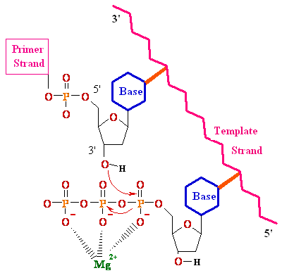
         Primer strand with free 3'- hydroxyl -OH group
         Template strand  (ss or ds with a nick)
         Deoxy-ribo-nucleoside 5'-tri-phosphates, dATP, dGTP, dTP and dCTP
 
          DNAn bases + dNTP  DNAn+1 + HO-O2P-O-PO-2OH
         Mg2+ to activate the dNTPs
 
         Enzyme catalyzes the nucleophilic attack of the 3'- hydroxyl on the alpha a phosphorous of the dNTP. Elongation proceeds in 5’--->3’ direction
         Enzyme is processive;  adds on dNTP after another and does not come off and on.
         Addition of dNTPs follows Watson-Crick base pairing rules
 
Mechanism of Nucleotide Addition


2. Proofreading activity of DNA polymerase
  DNA polymerases can also catalyze the hydrolysis of DNA strands that are unpaired or mismatches.
 
           3’--->5’ 3' nuclease activity examines each nucleotide added and removes mismatched residues before proceeding with polymerization
 
Error Probability =  Polymerization error (10-4)
                                                           X
                                   3'--->5' Nuclease error  (10-3)
                                   =  10-7   or  1 in 10,000,000
 
           5’®3’ nuclease activity results in the production of a free 5'-hydroxyl group and one or several dNMPs; responsible for removing RNA primers.
 
 
3. E. Coli Polymerase I Structure


E. Coli DNA Pol I, consist of three domains;  5’--->3’ exonuclease,  3’--->5’' exonuclease, and polymerase. Although Pol I is not the major replicative polymerase in E. Coli, it is the best studied DNA polymerase to date that exemplifies other DNA Pol enzymes.
36 kDa                                    67 kDa
 
           large cleft for duplex DNA
           flexible finger and thumb region for positioning of duplex and dNTPs
          polymerase site
           3’--->5’ and  5’--->3’ exonuclease catalytic sites
 
E. Coli DNA Polymerases
Characteristic Pol I Pol II Pol III
Mol. Weight (Da) 103 000 88 000 900 000
Number of polypeptides 1 4 10
rate (nucleotides / sec) 16-20 7 250-1000
  3’--->5’ exonuclease yes yes yes
 5’--->3’ exonuclease yes no no
function Primer removal,
gap filling
unknown Major replicative
polymerase
 
The study of DNA polymerases has lead to the discovery of :
                       anti tumor agents; i.e. Ara-C used against leukemia
                       anti viral agents; i.e. Acyclovir used against herpes simplex
polymerase chain reaction (PCR)
                                                which is used in all amplification reactions for cloning small amounts of DNA
 
V. DNA Replication
 
Because DNA is a complementary duplex, DNA replication must occur along both strands and it must proceed  5’®3’ on both strands. The DNA strands must be unwound and separated.  Several enzymes and DNA binding proteins are involved in initiating and maintaining DNA replication.
 
1.  Origin of replication
 
DNA replication begins at a specific site.  One such site in bacteria is the oriC site from E. coli.  Any circular DNA containing this sequence will be replicated just like the genome.
 
                       245 bp out of 4 000 000 bp
                       contains a tandem array the three 3 copies of 13-mers; GACNN
                       GAC common motif in oriC
                       A bp are common (facilitate unwinding)
 
2. Initiation of DNA replication in E. Coli: sequence of events
 
1.   dnaA recognizes the replication origin (oriC) and unwinds it at several sites
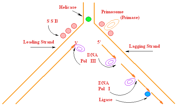
2. Helicase (dnaB) catalyzes ATP driven unwinding of ds DNA
3. DNA gyrase generates (-) super coiling that compensates for the (+) super coiling generated in front of the replication fork
4. Single stranded binding proteins (SSB) stabilizes unwound ssDNA
5.   3' ————————————————5'
                                 || Primase
      3' ————————————————5'
       5'••••••>
                                 || DNA Pol III 
      3' ————————————————5'
       5'••••••------------------------------------> 3'
                                 || DNA  Pol  I 
      3' ————————————————5'
                  5'----------------------------------------> 3'
 
DNA polymerase is primed by a small RNA strand which is produced by Primase, alpha RNA polymerase.
                  This is necessary because the polymerase can only add to a pre-existing 3'- hydroxy -OH.
 
3. Processive Synthesis
 
The synthesis of DNA in E. Coli is carried out by the DNA polymerase III holoenzyme (multi-subunit).  The synthesis is characterized by:
• Extremely high processivity; once it combines with the DNA and starts synthesis,
it does not come off the template.
• Tremendous catalytic potential: up to 1000 nucleotides per / sec.
• Error probability of 1 in 10 000 000 000; very high fidelity
• Contains the following three subunits:
- polymerase ( subunit)
- 3’--->5’ exonuclease ( subunit)
- a tight clamp for sliding DNA through ( subunit)
• Both leading and lagging strand are synthesized simultaneously.
Leading strand requires only one 1 primer. Lagging strand is synthesized discontinuously
(Okazaki fragments) and requires a new primer for each 1000-2000 nucleotides.
Primers are removed by 5’--->3’ activity of Pol I which also fills the gaps
Nicks are sealed by DNA ligase
                                                                                            The Replication Fork

               1. DNA Damage and Repair
 
B.      Types of DNA Mutations
 
Although the rate of mis-incorporation is very low (i.e. E. coli and Drosophila have mutation rates of
10-10, errors can occur due to the chemical nature of DNA bases and environmental factors.
 
1. Substitution of one base pair for another, A= for G=C, for example
            • most common form of mutation
            transition; G for A or  for C
 
                                     
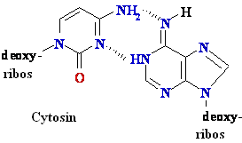
                                       Rare tautomer of Adenine
transitions occur when rare tautomers are present
 
trans-versions; G for  or C,          A for  or C,             for G or A,            C for G or A
 
2. Deletion of one 1 or more >1 base pairs
3. Insertion of one 1 or more >1 base pairs
 
1. Chemical Mutagens
 
Mutations can occur when the normal bases that are incorporated are changed.
 
2. Base analogs or bases that have altered hydrogen bonding capabilities can cause transitions.
            • ex. Bromo-uracil and Guanine or 2-amino-purine and cytosine
3.         Bases can be modified on the DNA by mutagens.
adenine is oxidatively de-aminated to hypoxanthine, cytosine to uracil, guanine to xanthine
4. Intercalating Agents
            insertion and deletion mutants
 
2. DNA Damage
  Base pairs can be damaged by environment or chemical factors, such as UV ~light or nitro-soureas.
  1. Pyrimidine dimers due to UV ~ light
 repaired by excision of the region by uvrABC exci-nuclease followed by gap filling by DNA Pol I.
            • can be repaired by photo-lyase, which splits the dimers
            • cause of xero derma pigmentosum, don't have exci-nuclease
 2. Nitro-soureas and other alkylating agents are obtained from smoking and other chemical contaminants.
  • result in methylation of G to form O6-Methyl G, which base pairs with  and
 to form  , which base pairs with G
            • there are some de-methylation enzymes
  3.         Cytosine deamination to
            spontaneous
           uracil-DNA glycosidase, AP exonuclease, DNA Pol I.
 4.         DNA repair is based on methylation of parental strand and a mismatch repair system that replaces the incorrectly incorporated base.